

Joseph R. Anticaglia, MD
Medical Advisory board
Low back pain (LBP) is common, costly, and treatable. It’s a common reason patients visit their family doctor, a common cause of disability, and costs Americans billions of dollars annually to manage this condition. Accurate diagnoses, and proper treatments are keys to successful outcomes.
Katherine, a 63 year old newspaper writer, left her Rheumatologist office after a routine examination because of her history of “weak, brittle bones,” osteoporosis. After leaving the doctor’s office, she entered a nearby coffee shop. She walked up to the counter, was about to order when a familiar voice called out to her. She turned, lost her balance, fell, and landed on her back. Workers behind the counter ran to her side, and helped her get up.
“Are you all right? Do you have any pain? Should we call 911?
“No, no. I’m OK. I’ll be fine.’
She walked out of the coffee shop to the corner of the street, flagged a taxicab, got home, and had lunch in her apartment. She was pain-free that afternoon, but in the early evening the low back pain became so intense she had to go to the ED, emergency department, for treatment.
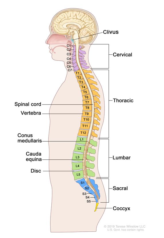
X-rays of the spine in the ED revealed she had compressed fractures of the lower spine, lumbar spine at levels L3, and L4 which were pinching the spinal nerves causing her severe pain.
She was examined in the ED by an Orthopedic spine surgeon who noted her medical history, examined her, and reviewed the X-rays of the spine with Kathrine. The surgeon pointed to the fractured bones on the X-rays, and said:
“The bones are pinching the nerves in the lower back causing your pain. “It looks like you may need surgery to relieve the pressure on the nerves caused by the broken bones. There are a lot of steps we need to take before we decide if it’s necessary to proceed with surgery.’
First, I’m going to prescribe medications to relax the muscles that have gone into spasm, and medications to relieve the pain. Please call my office, and make an appointment to see me in one week. We’ll go over in more detail the tests that are needed, and different treatment options. Right now, let’s make you as comfortable as possible. Here’s my number, call me, at any time if you have questions. You must take the pain medication” And that’s how Kathrine’s medical journey post-ED began.

The trip home from the ED was more than a “bumpy ride.” It was excruciating. It was worse than the ride to the ED. The slightest bump in the rode caused stabbing, knife-like pain. She couldn’t get comfortable in bed that night, didn’t sleep for three nights, and cancelled her one week doctor’s appointment.
Three weeks after the ED visit, Katherine hobbled into the spine surgeons office using a walker, and sat down. The doctor explained the various tests that had to be done to precisely diagnose the type, and extent of her injury and treatment choices.
The medical history and physical examination help determine the intensity and character of the pain (dull or sharp), and whether the pain is chronic lasting more than three months, or acute, lasting less than six weeks. In addition, the H&P gives information about the location of the pain, for example, is it located solely in the lower back, or does the pain radiate down your legs , or to other parts of the body? Do you have a fever? Any problems with bowel movement, or trouble urinating? Do you have pain at rest, night pain?
Image studies can demonstrate if there are broken bones, arthritis, spinal stenosis, bulging discs, and whether there’s involvement of the muscles, tendons, or ligaments.
Magnetic resonance imaging (MRI) uses a powerful magnet, radio waves and a computer to make detailed images of the spinal cord and spinal nerves. Katherine had a standing, “upright” MRI of the spinal column which showed more detail of the soft tissue structure of the spinal column.
Computed tomography (CT) scan uses X-rays , and computers to produce images which are particularly helpful in demonstrating the bony structures of the spinal column. She also had a CT scan done which helped doctors pinpoint more accurately the bony fractures.
X-rays create pictures of bones and soft tissues that can show fractures, as in Katherine’s case when she was in the ED. Katherine’s blood test revealed no evidence of an infection, or other problems.
Electromyogram (EMG) and myelogram were not recommended in Katherine’s situation (see glossary).
Low back pain usually gets better after a few days of rest, and the majority of patients will be symptom free in about four weeks with non-surgical management. Heat, ice, moderate exercise, and over the counter pain medication offer temporary pain relief. Patients are encouraged to return to normal activity as soon as feasible. Patient discussions should include proper lifting techniques, weight loss, and the advantages of exercising, and physical therapy.
Physical therapists can design a personalized exercise program that can improve your flexibility, and strengthen back muscles. Your doctor may prescribe medications to relieve your pain, or muscle relaxants to prevent back spasms. Often, doctors initially do not recommend PT for Kathrine-like problems.
Katherine’s last medical journey to relieve her back pain involved steroid injections into the area of the lower back that was causing her pain. She agreed to two injections, both of which were unsuccessful. What’s next?
When Katherine entered her surgeon’s office for the pre-operative visit, she was surprised that the wall was plastered with all of her x-rays, and image studies. He reviewed the films, laboratory, and consultation reports. He described the surgery she was scheduled to undergo,
“I’m going to remove pieces of bone from the lamina, the back part of the involved bones of the lower spine to relieve pressure on the nerves that are causing your pain.” The surgery was a success, and Katherine was surprised that hours after the operation, the nurses insisted she get up from bed, take a few steps. She was told that walking and swimming were the best PT exercises for her. The surgical journey was over, but her personal journey began with those first few steps.
Electromyography (EMG) measures how fast an electrical impulse moves through your nerve. It tests for nerve damage that can cause numbness, and tingling in your legs
A myelogram is a procedure that combines the use of x rays or CT scans with a contrast dye that is injected into the spinal column to look for lower back problems.
NIH tips to strengthen lower back and abdominal muscles.
Exercise regularly to keep your muscles strong and flexible. Consult a physician for a list of low-impact, age-appropriate exercises that are specifically targeted to strengthening lower back and abdominal muscles.
Maintain a healthy weight and eat a nutritious diet that promotes new bone growth.
Use ergonomically designed furniture and equipment at home and at work.
Switch sitting positions often and periodically walk around the office or gently stretch your muscles to relieve tension.
Put your feet on a low stool or a stack of books when sitting for a long time.
Wear comfortable, low-heeled shoes.
Sleeping on your side with your knees drawn up in a fetal position can help open up the joints in the spine and relieve pressure by reducing the curvature of the spine. Always sleep on a firm surface.
Don’t try to lift objects that are too heavy. Lift from the knees, keep a straight back, and objects close to the body.
Quit smoking. Smoking reduces blood flow to the lower spine, which can contribute to spinal disc degeneration. Smoking also increases the risk of osteoporosis and impedes healing. Coughing due to heavy smoking also may cause back pain.
This article is intended solely as a learning experience. Please consult your physician for diagnostic and treatment options.