

Joseph R. Anticaglia, MD
Medical Advisory board
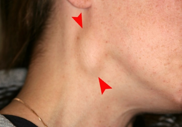
An anxious mother takes her child to the Pediatrician concerned about a bump that appeared “out of nowhere” on one the side of the neck. Another mother is worried about swollen lymph nodes on both sides of her child’s neck that haven’t gone away after two months. A nervous Mom brings her child to the doctor because she’s worried about a lump in the midline of the neck above the Adam’s Apple (thyroid cartilage) after the child had a cold. What causes these bumps and lumps? How serious are they? When should a parent worry?
There are three main causes of neck masses in children.

Bumps or lumps in the neck are quite common in infants, and children. The majority of neck masses in children, particularly if noticed on both sides of the neck, are benign lymph nodes that become enlarged after an upper respiratory infection (URI). The enlarged lymph nodes are commonly caused by a cold or sinus infection. Ear infections, and sore throats are other common causes of enlarged nodes.
After a bacterial or viral URI, lymph nodes in the neck can become enlarged in their attempt to fight the infection. The infection can cause a spike in the child’s temperature, and the lymph nodes to become red and painful to the touch. The enlarged lymph nodes can persist without pain for several months after the infection has been successfully treated with antibiotics, or after the viral infection has cleared. One needs to rule out others causes of infectious enlargement of lymph nodes in children, such as cat scratch fever, tick bites, and mycobacterial infections.
Congenital bumps or lumps are present at birth due to abnormal embryonic development. The location of the neck mass is useful in diagnosing congenital neck masses. For instance, the most common “midline” neck mass in children is a thyroglossal duct cyst. It’s usually diagnosed in the first five years of life; the cyst moves up and down when the child swallows, and can become infected.
The second most common congenital neck mass in children is a branchial cleft cyst. These abnormal developmental cysts are most commonly located on the side of the neck in its upper one third. Of the congenital neck masses, approximately 70% are thyroglossal duct cysts, and 25% branchial cleft cysts. Vascular abnormalities may or not be present at birth.
Cancer is the second most common cause of deaths in children over five years of age after deaths due to accidental trauma. A one sided neck mass in children on rare occasions can be caused by a non-Hodgkin’s lymphoma or Hodgkin’s lymphoma.
Lymphocytes are white blood cells in the immune system that protect us by fighting off infections and foreign bodies. If these cells multiply uncontrollably, they can form a type of cancer called lymphoma.
Lymphomas, according to a U. of Chicago report, “accounts for half of pediatric head and neck malignancy, with non-Hodgkin’s lymphoma in 60% of cases and Hodgkin’s lymphoma in 40%.”
Hodgkin’s lymphoma is a curable malignant disease that causes a painless enlargement of the lymph nodes, liver, and spleen.
What is particularly worrisome in children with a neck mass is the finding that the mass is located on one side of the neck, larger than one inch, has a hard or rubbery consistency, fixed in place with no signs of inflammation, and not responding to antibiotics.
A neck mass with a of a URI, fever, redness, and pain suggest a diagnosis of an infection, or inflammation. A neck mass present at birth makes one investigate a congenital/developmental cause for the mass. A painless mass, larger than one inch that’s hard which doesn’t move, and is located on one side of the neck needs further evaluation to rule out a malignancy
A doctor may order a complete blood count (CBC) to determine whether an infection is the reason for the swollen lymph nodes. Often, the initial image study ordered by a doctor is an ultrasound which uses sound waves to create images of the neck and lymph nodes. Is it a solid, or cystic mass?
An MRI scan uses a magnetic field and radio waves to generate computerized images of the body. A CT scan (computerized tomography) uses X-rays and a computer to create images of the body. A biopsy is recommended if the imaging tests suggest (as well as the medical history, physical findings and laboratory tests suggest) the neck mass may be a tumor. The small tissue sample obtained from the biopsy is sent to the laboratory for pathologic diagnosis.
Treatment depends on the medical history, physical examination, characteristics of neck mass, and diagnostic tests. If the neck mass is caused by a virus, or bacterial infection, appropriate medications are prescribed. If the neck mass remains enlarged after treatment, depending on the medical history and findings, your doctor may suggest observation.
If an ultrasound of the neck demonstrates pus in the neck mass, he or she may recommend an incision and drainage. Your doctor may advise surgery if there is a congenital malformation, such as a thyroglossal duct cyst. If a biopsy of the neck is cancerous, treatment is based on the type of cancer.
Although the vast majority of pediatric neck masses are non-cancerous, consult your pediatrician if you have any questions about lumps, or bumps in your child’s neck.
Lymphadenitis is an infection in one or more lymph nodes which can become enlarged, red, or tender.
B Symptoms are found in lymphomas, and refer to fever, drenching night sweats, and loss of more than 10% of body weight over six months.
Mycobacterium is a group of bacteria, mycobacteria that can cause neck masses. It includes the causative agents of tuberculosis (TB) and leprosy.
This article is intended solely as a learning experience. Please consult your physician for diagnostic and treatment options.